White Spots on Teeth: Causes and What to Do
The two common reasons enamel turns chalky white, and the honest guide to which spots you can rebuild at home and which need professional care.
- Most white spots on teeth come from one of two things: early demineralization, where acid has drawn mineral out of the surface, or dental fluorosis, a cosmetic change formed while the teeth were developing.
- An early demineralization spot is the reversible kind. Because the surface is still intact, the mineral underneath can be rebuilt with fluoride or hydroxyapatite and better daily habits.
- Fluorosis spots are baked into the enamel from childhood and will not remineralize away; they are a cosmetic finish, not active decay, and are managed by a dentist if appearance matters.
- You cannot reliably tell an arrestable white spot from one that has started to cavitate by eye. A spot that is turning brown, feels rough, or is sensitive should be seen in person.
- White spots are a warning worth acting on early. Caught while the surface is intact, many are genuinely arrestable; left to progress, they can become cavities that no home routine can fix.
White spots on teeth are usually either early demineralization, where acid has leached mineral from the surface, or fluorosis, a cosmetic change from childhood tooth development. Demineralization spots can often be remineralized with fluoride or hydroxyapatite and better habits while the surface is intact; fluorosis will not fade and is managed cosmetically. Any spreading, brown, or sensitive spot needs a dentist.
Why enamel turns chalky white
A white spot is an optical signal, not a stain. Healthy enamel is a dense, glassy crystal that light passes through smoothly. When acid dissolves mineral out of the layers just beneath the surface, it leaves microscopic pores; light scatters off those pores instead of passing through, and the patch looks chalky and matte, especially when the tooth is dried. This is the early caries lesion, a biofilm-mediated, sugar-driven demineralization of the surface, and crucially the outer skin of enamel is often still intact over it. That intact surface is what makes the spot reversible: fluoride, hydroxyapatite and salivary calcium and phosphate can diffuse in and rebuild the mineral, and the pores shrink until light passes through normally again. Fluorosis produces a similar chalky look but by a completely different route. It forms while the enamel is being laid down in childhood, when higher fluoride exposure alters how the crystal matures, leaving a porous, mottled finish that is set for life. Both look white; only one is an active, rebuildable process.
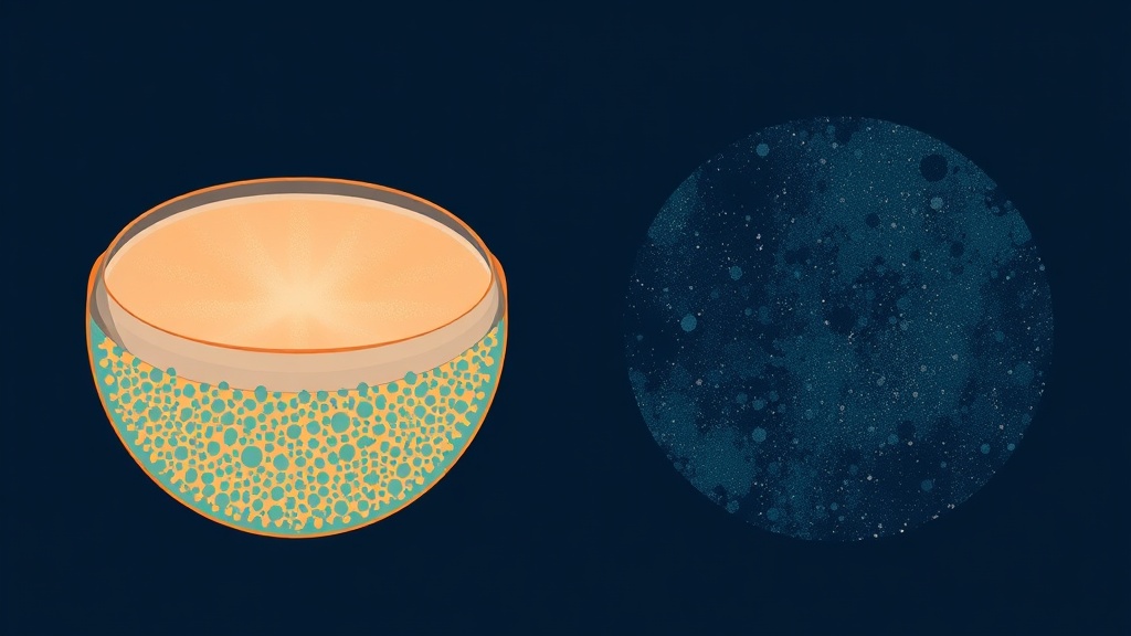
Both look chalky, but a demineralization spot is a rebuildable porous layer under an intact surface, while fluorosis is a cosmetic finish set during tooth development.
What the research actually shows
Every claim below maps to a named, peer-reviewed source in the Sources section. According to PubMed.
| Claim | Evidence | Source |
|---|---|---|
| Dental caries is a biofilm-mediated, sugar-driven disease that causes phasic demineralization and remineralization of the tooth surface; the white spot is its earliest visible stage. | Consensus disease-definition primer. | Pitts et al., 2017 |
| An active early lesion is progressively demineralizing while an arrested lesion is progressively remineralizing; activity, not just presence, is what a clinician assesses. | Study of caries lesion activity. | Hoxie et al., 2023 |
| Many lesions that looked advanced on X-ray were still non-cavitated after tooth separation, 79.6 percent overall, meaning many are candidates for remineralization rather than drilling. | Clinical study using tooth separation. | Nascimento et al., 2020 |
| Around 12 percent of people had fluorosis of aesthetic concern at 0.7 ppm water fluoride; most fluorosis is a cosmetic change rather than a disease. | Cochrane review of water fluoridation. | Iheozor-Ejiofor et al., 2024 |
| Resin infiltration of early proximal lesions cut 18-month progression to 7 percent versus 37 percent in untreated controls, a dentist-applied option for early white spots. | Randomized controlled trial. | Paris et al., 2010 |
Telling white spots apart
| Type of white spot | What causes it | What to do |
|---|---|---|
| Early demineralization (active) | Recent acid attacks near plaque, braces or the gumline | Remineralize: fluoride or hydroxyapatite, lower sugar frequency, better cleaning |
| Arrested demineralization | An old lesion that has already re-hardened | Usually leave it; keep up good habits and monitor |
| Dental fluorosis | Higher fluoride exposure while teeth were forming in childhood | Cosmetic only; a dentist can address appearance if it bothers you |
| Early cavitation (broken surface) | A demineralized spot that has progressed to a hole | See a dentist; this cannot be remineralized at home |
Where white spots come from, and the ones to watch
Active demineralization spots tend to appear exactly where plaque sits undisturbed: along the gumline, between teeth, and famously around orthodontic brackets, where they can show up as bright squares after braces come off. That location is a clue, because it points to a spot you can influence by cleaning better and cutting acid exposure. The reassuring finding from careful clinical studies is that a large share of lesions that look worrying, even on X-ray, are still non-cavitated and therefore arrestable, so a white spot is far from an automatic filling. The catch is that the earliest lesions are also the hardest to judge; even trained examiners misclassify them, which is why activity and depth are a clinician''s call rather than a mirror-and-phone-light call. The spots to take seriously are the ones changing: a patch that is enlarging, picking up a brown tinge, developing a rough or catchy surface, or becoming sensitive. Those signs suggest the process is winning, and at that point the honest answer is professional assessment, not a longer home routine.
Evidence you can act on.
Occasional emails — new research, new protocols, no noise.
What to do about a white spot
This supports the enamel surface and is aimed at early, non-cavitated demineralization spots on sound teeth. It does not treat decay as a disease, and fluorosis will not respond to it because it is not active.
- 1
Get the spot staged by a dentist first
as soon as practicalBefore you commit to a routine, have a professional tell you whether the spot is active demineralization, arrested, fluorosis, or already cavitated. That single answer decides everything that follows, and it is not reliably a home judgement.
- 2
Clean the exact spot better
twice dailyActive spots live where plaque sits. Angle your brushing to the gumline, clean between the teeth daily, and pay particular attention around any brackets or crowded areas so the surface is not constantly re-acidified.
- 3
Brush with fluoride or nano-hydroxyapatite and leave a film
2 minutes, twice dailyBoth ingredients rebuild early lesions; spit rather than rinse so a thin mineral layer stays on the surface. For stubborn spots a dentist may add professional-strength fluoride to the specific tooth.
- 4
Lower the frequency of acid attacks
every dayCluster sugary and acidic foods and drinks into fewer moments and keep them away from bedtime. Fewer acid dips give the spot longer rebuilding windows, which is what shifts an active lesion toward arrest.
- 5
Ask about cosmetic options for what will not fade
as neededIf a spot is fluorosis or an arrested scar that bothers you cosmetically, a dentist has options such as resin infiltration or micro-finishing. These are appearance treatments, not remineralization, and belong in professional hands.
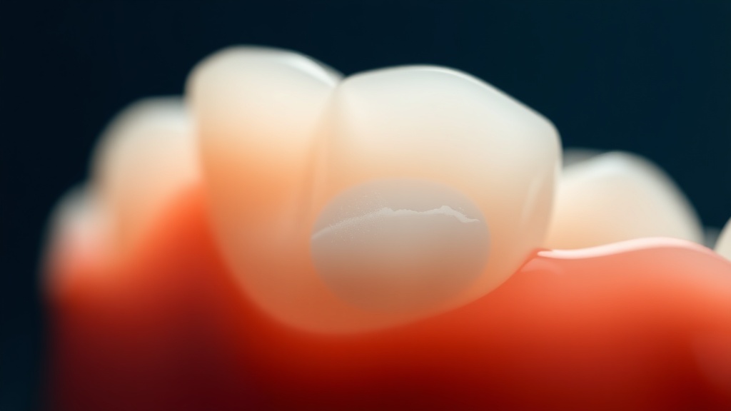
Active white spots cluster where plaque sits, at the gumline, between teeth and around brackets, which is also where better cleaning does the most good.
See a dentist if a white spot is enlarging, turning brown, feels rough or catchy, or has become sensitive, or if you can feel any softness or a hole with your tongue or floss; those suggest the lesion may be progressing beyond what home care can reach. It is also worth an in-person visit simply to confirm what a spot is, since an arrestable early lesion and a cavitated one can look alike, and fluorosis needs cosmetic rather than remineralizing care.
Frequently asked questions
Sources
- 1.
- 2.
- 3.
- 4.
- 5.
- 6.
- 7.
- 8.
- 9.

Fix your breath at the source.
The complete science-backed protocol — engineered to eliminate volatile sulfur compounds at the biological source.
Start the Breath Protocol →Related reading
Educational purposes only. The content on this page is not medical advice and is not a substitute for consultation with a qualified dental or medical professional.
More from the library
 Answers8 min
How to Remineralize Teeth: An Honest, Practical Guide
The practical routine that supports enamel remineralization, and the clear line between the early lesions it can rebuild and the decay it cannot.
Read →→ Ingredients8 min
How Fluoride Toothpaste Remineralizes Teeth
The chemistry behind fluoride's remineralizing power, and the honest line between the early lesions it can rebuild and the cavities it cannot.
Read →→ Guides8 min
Does Hydroxyapatite Toothpaste Work? What the Evidence Shows
The fluoride-free ingredient dentists keep debating, measured against the actual clinical trials.
Read →→ Comparisons8 min
Hydroxyapatite vs Fluoride: Which Toothpaste Is Right for You?
Two ways to strengthen enamel, one big difference in how much evidence stands behind each.
Read →→ Answers8 min
How to Get Rid of White Spots on Teeth: Your Options, Ranked
An honest, evidence-based ladder of options, from what you can do at home to what only a dentist can fix.
Read →→ Ingredients8 min
Hydroxyapatite Toothpaste Benefits: What the Evidence Actually Supports
A calm, science-first look at what hydroxyapatite toothpaste can genuinely do for your enamel, and where the evidence stops.
Read →→