
White Spot Lesions on Teeth
What a white spot lesion actually is, how it differs from a cavity, and the active-versus-arrested distinction that decides what happens next.
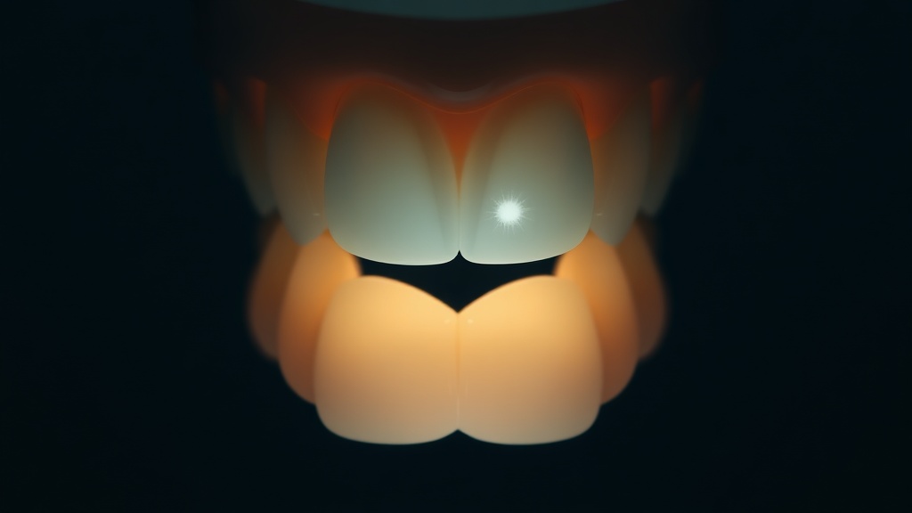
- A white spot lesion is the earliest visible stage of tooth decay: acid has demineralized the enamel just beneath an intact surface, making it porous and chalky, but no hole has formed yet.
- It is defined by an intact surface over a softened core - that is exactly why it can still be reversed, and exactly why it is fragile: if the surface breaks and the lesion cavitates, it is past the point of home repair.
- The classic cause is orthodontic decalcification: plaque trapped around brackets bathes the enamel in acid, so white spot lesions appear as halos when braces come off.
- Clinicians distinguish active lesions (chalky, matte, rough - progressively demineralizing) from arrested lesions (shiny, harder - progressively remineralizing); the goal of care is to flip an active lesion to arrested.
- Many lesions that look advanced on an X-ray are still non-cavitated and genuinely arrestable, which is why staging by a dentist - not drilling on sight - is the modern standard.
A white spot lesion is subsurface enamel that has lost mineral to acid while its outer surface stays intact - the first visible stage of a cavity. Because the surface has not broken, the lost mineral can often be partly redeposited, so an early, active lesion can be arrested and remineralized rather than drilled, if it is caught in time.
What a white spot lesion actually is
To understand a white spot lesion you have to picture the enamel in cross-section. Dental caries is a biofilm-mediated, sugar-driven, dynamic disease in which the enamel constantly swings between losing and regaining mineral. When acid from plaque wins that tug-of-war, it does something counterintuitive: it dissolves mineral from the layer just below the surface while leaving a relatively intact surface skin on top. The result is a porous subsurface zone with a still-solid roof. Light travelling into that porous enamel scatters instead of passing cleanly through, so the spot looks chalky, opaque and white - hence the name. This subsurface-with-intact-surface architecture is the whole story, because it explains both the hope and the fragility. The hope: mineral can diffuse back in through the intact surface and rebuild the crystals, so an early lesion is genuinely reversible. The fragility: that surface roof is thin, and once it collapses into an actual cavity, the enamel - which is acellular and cannot regenerate itself - can no longer be rebuilt at home. A white spot lesion is therefore best understood as a cavity that has not happened yet.

The defining feature: a porous, demineralized subsurface zone sealed under a still-intact enamel surface.
What the research actually shows
Every claim below maps to a named, peer-reviewed source in the Sources section. According to PubMed.
| Claim | Evidence | Source |
|---|---|---|
| Caries is a biofilm-mediated, sugar-driven, dynamic disease of phasic demineralization and remineralization of the dental hard tissues. | Nature Reviews Disease Primers consensus. | Pitts et al., 2017 |
| An active lesion is progressively demineralizing while an arrested lesion is progressively remineralizing - the clinical distinction that guides care. | Review of caries lesion activity assessment. | Hoxie et al., 2023 |
| A lesion can be arrested at any stage provided clinically plaque-free conditions are obtained. | Foundational clinical cariology review. | Nyvad & Fejerskov, 1997 |
| 79.6% of lesions that looked advanced on X-ray were still non-cavitated after tooth separation - candidates for arrest, not drilling. | Clinical study using temporary tooth separation. | Nascimento et al., 2020 |
| Resin infiltration cut 7-year proximal lesion progression to 9% versus 45% in untreated controls. | Long-term randomized controlled trial. | Paris et al., 2020 |
Active versus arrested lesions
| Feature | Active lesion | Arrested lesion |
|---|---|---|
| Surface look | Chalky, matte, opaque | Shinier, glossier |
| Texture | Rough to a probe | Smoother, harder |
| Direction | Progressively demineralizing | Progressively remineralizing |
| What it needs | Plaque control and remineralization now | Monitoring to keep it stable |
| Home repair possible? | Yes, if surface still intact | Already stabilized |
Why braces are the classic trigger
If there is a signature cause of white spot lesions, it is orthodontic treatment. Brackets and wires create dozens of sheltered corners where a toothbrush struggles to reach, so plaque accumulates and stays put against the enamel around each bracket. That plaque generates acid, the acid demineralizes the enamel in a ring, and when the braces finally come off the lesions are revealed as pale halos framing where each bracket sat - a pattern clinicians call decalcification. The frustrating part for patients is that these appear precisely on the highly visible front teeth that braces were meant to improve. The reassuring part is that a decalcification lesion is still just a white spot lesion, subject to the same rules as any other: caught early, with meticulous plaque removal and remineralizing products, an active lesion can be nudged toward arrest, and its appearance can be improved. Left to its own devices, though, it can progress or simply persist as a stubborn cosmetic mark - which is why orthodontic patients are counselled so heavily on hygiene, and why a post-braces white spot is worth acting on rather than ignoring.
Evidence you can act on.
Occasional emails — new research, new protocols, no noise.
Managing an early white spot lesion
If a dentist has identified an early, non-cavitated lesion, the aim is to flip it from active to arrested. These steps support that; none of them is a substitute for professional staging.
- 1
Get the lesion staged and its activity assessed
one visitA dentist checks whether the surface is intact and whether the lesion looks active (chalky, rough) or arrested (shiny, hard). That judgement decides everything that follows.
- 2
Remove plaque from the lesion meticulously
twice dailyArrest depends on keeping the surface as plaque-free as possible, since the biofilm is the acid source. For braces-related spots, that means targeted cleaning around every bracket with interdental aids.
- 3
Remineralize with fluoride or hydroxyapatite
twice dailyA 1,000-1,500 ppm fluoride or a hydroxyapatite toothpaste supplies mineral to redeposit through the intact surface; spit, do not rinse. A nightly leave-on layer adds contact time.
- 4
Ask about resin infiltration for stubborn lesions
discuss at visitFor lesions that will not remineralize or that bother you cosmetically, a dentist can infiltrate them with a no-drill resin that both stabilizes and visually blends the spot; trials show durable control of early lesions.
- 5
Re-check on a schedule
as advisedBecause activity can change, lesions are monitored over time rather than treated once and forgotten. Many that look concerning are still arrestable, so patient follow-up beats premature drilling.
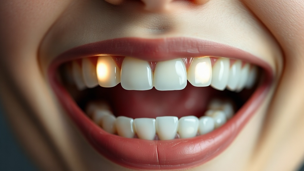
Orthodontic decalcification leaves pale halos framing where each bracket sat - the classic white spot lesion pattern.
Any white spot lesion should ideally be assessed in person, because the single decision that matters most - is the surface still intact, or has it cavitated - cannot be made reliably at home. See a dentist promptly if a spot feels rough or catches a fingernail, is darkening, appears during or after orthodontic treatment, or comes with sensitivity. Early, active lesions are exactly the cases where prompt professional care can arrest the process without any drilling.
Frequently asked questions
Sources
- 1.
- 2.
- 3.
- 4.
- 5.
- 6.
- 7.

Fix your breath at the source.
The complete science-backed protocol — engineered to eliminate volatile sulfur compounds at the biological source.
Start the Breath Protocol →Related reading
Educational purposes only. The content on this page is not medical advice and is not a substitute for consultation with a qualified dental or medical professional.
More from the library
 Causes8 min
What Causes White Spots on Teeth? The Honest Differential
The four common causes, how each one forms, and how to tell which kind you are looking at.
Read →→ Answers8 min
How to Get Rid of White Spots on Teeth: Your Options, Ranked
An honest, evidence-based ladder of options, from what you can do at home to what only a dentist can fix.
Read →→ Guides8 min
White Spots on Teeth: Causes and What to Do
The two common reasons enamel turns chalky white, and the honest guide to which spots you can rebuild at home and which need professional care.
Read →→ Ingredients8 min
How Fluoride Toothpaste Remineralizes Teeth
The chemistry behind fluoride's remineralizing power, and the honest line between the early lesions it can rebuild and the cavities it cannot.
Read →→ Ingredients8 min
Hydroxyapatite Toothpaste Benefits: What the Evidence Actually Supports
A calm, science-first look at what hydroxyapatite toothpaste can genuinely do for your enamel, and where the evidence stops.
Read →→ Answers8 min
Can You Rebuild Tooth Enamel? What Is Actually Possible
The honest split: what you can genuinely strengthen at home, and what only a dentist can restore.
Read →→