What Causes White Spots on Teeth?
The four common causes, how each one forms, and how to tell which kind you are looking at.
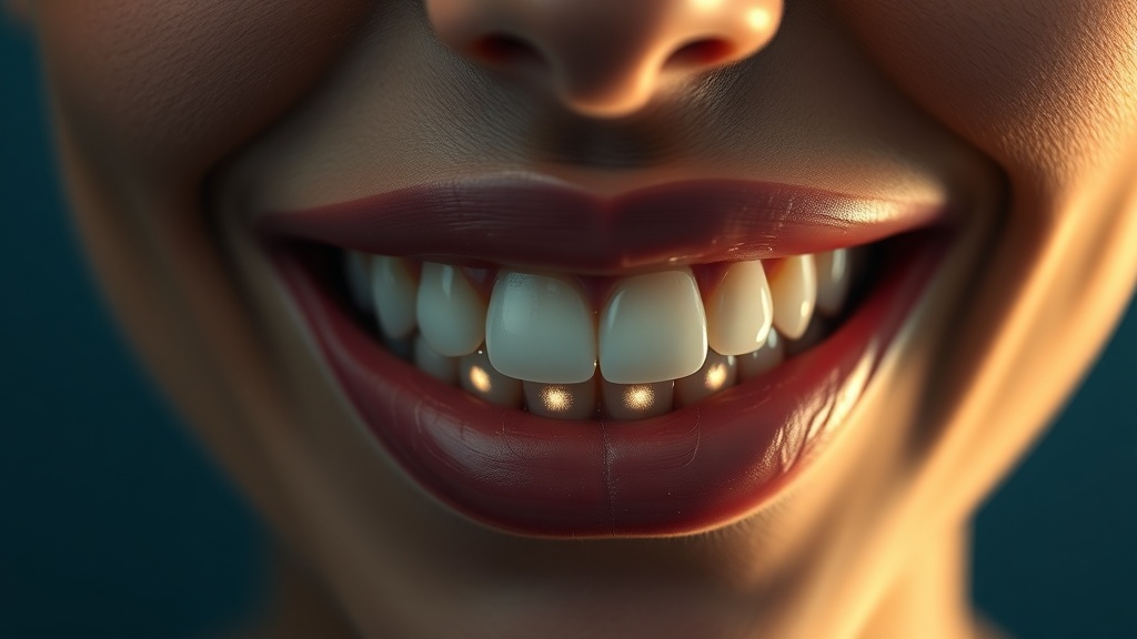
- Most white spots come down to four causes: early enamel demineralization, dental fluorosis, enamel hypoplasia, and temporary dehydration - and they form in very different ways.
- Demineralization spots form now, from acid pulling mineral out of the enamel surface; the classic examples are plaque left along the gumline and the marks that appear around braces brackets.
- Fluorosis and hypoplasia both form years earlier, while the tooth is still developing under the gum, so they are set into the enamel by the time the tooth erupts - not something you caused this month.
- The pattern is the biggest clue: demineralization tends to sit where plaque collects, fluorosis is usually diffuse and symmetrical across matching teeth, and hypoplasia often shows as a defined pit or band on specific teeth.
- Telling them apart matters because one of them - active demineralization - is an early cavity in the making, while the others are cosmetic; only an in-person exam can stage a spot reliably.
White spots usually come from one of four things: early demineralization (acid drawing mineral out of the enamel, as around plaque or braces), dental fluorosis (too much fluoride while teeth formed), enamel hypoplasia (enamel that formed thin or defective from illness or nutrition), or simple dehydration of the enamel. Only the first is an active process you can still influence.
Demineralization: the spot that is forming right now
This is the only cause on the list that is happening in real time, and it is the one to take seriously. Every time you eat, plaque bacteria turn sugars into acid, and when the pH at the tooth surface drops below roughly 5.5 - the so-called critical pH - the enamel begins to dissolve, losing calcium and phosphate from just beneath the surface. Do that often enough without letting the tooth recover, and a chalky, opaque patch appears where the enamel has gone porous. It is not random where these show up: they cluster exactly where plaque is hardest to remove and sits longest - along the gumline, between teeth, and famously in a halo around orthodontic brackets, where they are called decalcification or white-spot lesions. Crucially, this early stage is still reversible in principle: as long as the surface has not broken into a cavity, the lost mineral can be partly redeposited. That is what makes an early demineralization spot both the most concerning cause and the most hopeful one - it is a warning that has not yet become a hole.
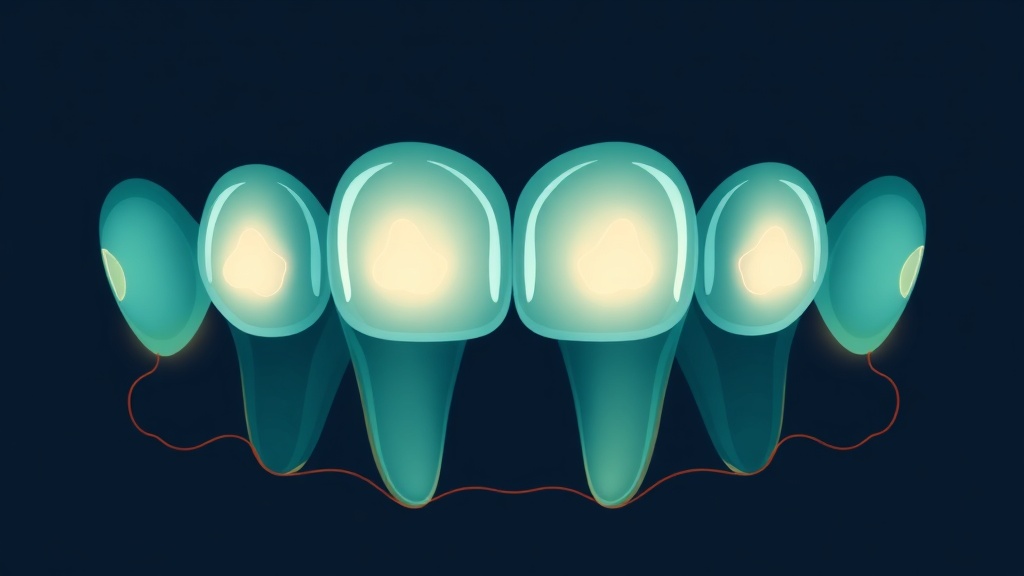
Demineralization spots cluster where plaque lingers longest - the gumline and the halo around braces brackets.
What the research actually shows
Every claim below maps to a named, peer-reviewed source in the Sources section. According to PubMed.
| Claim | Evidence | Source |
|---|---|---|
| Enamel begins to dissolve below a critical pH of about 5.5, which is why frequent acid exposure drives demineralization spots. | Review of erosion and demineralization thresholds. | Lussi et al., 2011 |
| About 12% of people had fluorosis of aesthetic concern at 0.7 ppm water fluoride, and roughly 40% had some detectable level. | Cochrane systematic review of water fluoridation. | Iheozor-Ejiofor et al., 2024 |
| Around 23% of US people aged 6-39 had at least very mild fluorosis, most of it cosmetic. | NHANES national surveillance data. | Beltran-Aguilar et al., 2005 |
| An early non-cavitated spot can still be arrested at any stage if plaque-free conditions are achieved. | Foundational clinical cariology review. | Nyvad & Fejerskov, 1997 |
| Once enamel cavitates it cannot repair itself, so a broken-surface spot is past the reversible stage. | Materials-science review of enamel repair. | Liu et al., 2022 |
Telling the four causes apart
| Cause | When it formed | Typical pattern | Still active? |
|---|---|---|---|
| Demineralization | Now, ongoing | Along the gumline, between teeth, around braces | Yes - can worsen or improve |
| Dental fluorosis | During tooth development | Diffuse, symmetrical across matching teeth | No - set in the enamel |
| Enamel hypoplasia | During tooth development | Defined pit, band or patch on specific teeth | No - a formed defect |
| Dehydration | Temporary | Whole tooth looks chalky, resolves in hours | No - not a true lesion |
Fluorosis and hypoplasia: spots that formed years ago
The other two common causes have nothing to do with how you brush today, because both were written into the enamel while the tooth was still forming under the gum in childhood. Dental fluorosis happens when a child takes in more fluoride than needed during those years - from swallowed toothpaste, high-fluoride water and supplements combined - which subtly disrupts how the enamel mineralizes, leaving diffuse white flecks or mottling. It is common and, at the levels seen with optimally fluoridated water, overwhelmingly a mild cosmetic finding rather than a health problem; the honest framing is that fluorosis is a reason some people prefer fluoride-free products, not evidence of poisoning. Enamel hypoplasia is different again: here the enamel-forming cells were disturbed by a high fever, a childhood illness, nutritional deficiency, premature birth or local trauma, so the enamel came in physically thin or defective. That is why hypoplasia often looks like a sharply defined patch, pit or horizontal band on particular teeth rather than a soft diffuse haze. Because both are finished structures, no toothpaste will remineralize them away - improving their appearance is a cosmetic job for a dentist.
Evidence you can act on.
Occasional emails — new research, new protocols, no noise.
How to read your own white spots
You cannot diagnose a spot at home, but you can gather good clues before your appointment. Work through these observations.
- 1
Look at where the spots sit
1 minuteSpots hugging the gumline, crowded between teeth, or ringed around old brace positions point toward demineralization. Spots spread evenly across several matching teeth point more toward fluorosis.
- 2
Check for symmetry
1 minuteFluorosis is typically symmetrical - similar marks on the same teeth on both sides - because both sides developed together. A lone, well-defined patch on one tooth leans toward hypoplasia or a single demineralized spot.
- 3
Notice the edges and texture
1 minuteA soft, hazy, blend-into-the-tooth edge suggests demineralization or fluorosis. A sharp border, a pit, or enamel that feels rough or thin suggests hypoplasia. Anything that catches a fingernail deserves prompt attention.
- 4
Watch after brushing and drying
a few hoursIf the whole tooth looks chalky right after drying but evens out within hours, that is simple dehydration, not a lasting spot - common after wearing a mouthguard or aligners.
- 5
Have it staged in person
one visitBring your observations to a dentist. The one outcome worth ruling out is an active early cavity masquerading as a cosmetic mark, and that judgement needs a trained eye and good light.
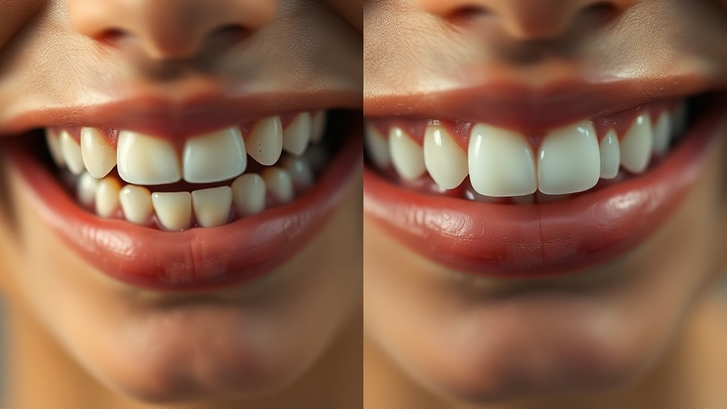
Diffuse and symmetrical points to fluorosis; a single sharp-edged patch points to hypoplasia or a localized lesion.
Book a dental exam if a white spot is new, feels rough or catches a fingernail, is darkening, or comes with sensitivity, because those features can signal active early decay rather than a cosmetic mark. New spots appearing during or just after orthodontic treatment are worth flagging quickly, since early decalcification responds best when caught fast. A dentist can confirm the cause, stage any active lesion, and explain cosmetic options for fluorosis or hypoplasia if their appearance bothers you.
Frequently asked questions
Sources
- 1.
- 2.
- 3.
- 4.
- 5.

Fix your breath at the source.
The complete science-backed protocol — engineered to eliminate volatile sulfur compounds at the biological source.
Start the Breath Protocol →Related reading
Educational purposes only. The content on this page is not medical advice and is not a substitute for consultation with a qualified dental or medical professional.
More from the library
 Guides8 min
White Spots on Teeth: Causes and What to Do
The two common reasons enamel turns chalky white, and the honest guide to which spots you can rebuild at home and which need professional care.
Read →→ Answers8 min
How to Get Rid of White Spots on Teeth: Your Options, Ranked
An honest, evidence-based ladder of options, from what you can do at home to what only a dentist can fix.
Read →→ Guides8 min
White Spot Lesions on Teeth: The Clinical Explainer
What a white spot lesion actually is, how it differs from a cavity, and the active-versus-arrested distinction that decides what happens next.
Read →→ Ingredients8 min
Remineralizing Toothpaste: How It Works and What Actually Rebuilds Enamel
A plain-English guide to how remineralizing toothpaste works — and an honest comparison of fluoride, n-HA and CPP-ACP.
Read →→ Ingredients8 min
Hydroxyapatite Toothpaste Benefits: What the Evidence Actually Supports
A calm, science-first look at what hydroxyapatite toothpaste can genuinely do for your enamel, and where the evidence stops.
Read →→ Answers8 min
Can You Rebuild Tooth Enamel? What Is Actually Possible
The honest split: what you can genuinely strengthen at home, and what only a dentist can restore.
Read →→