
Dental Fluorosis: Why Teeth Get White or Brown Spots
A calm, evidence-based look at what dental fluorosis is, why it forms only during childhood, and the cosmetic options that can improve how it looks.
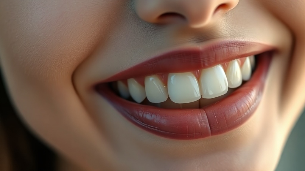
- Dental fluorosis is not a stain that lands on the tooth from outside — it forms inside the enamel while the tooth is still developing under the gum, so it is a permanent feature of how that enamel was built, not something you can brush or bleach off the surface.
- It is common and usually very mild: in recent US survey data most children and teens showed some degree of fluorosis, and the large majority of cases are the faint, barely-noticeable kind — small chalky-white flecks or lacy lines rather than obvious marks.
- It happens only during childhood tooth formation, roughly from birth to about age 8, when more fluoride was taken in than the developing enamel could handle; once teeth have erupted, fluoride can no longer cause or worsen it.
- Mild fluorosis shows up as white opaque spots or streaks; more severe fluorosis can leave pitting and brown mottling, because the porous enamel picks up colour later from food and drink.
- You cannot undo fluorosis, but you can improve how it looks: minimally-invasive cosmetic options like enamel microabrasion and resin infiltration are well studied, and moderate-to-severe cases should be assessed in person by a dentist.
Dental fluorosis is a cosmetic change in enamel that forms when a child takes in more fluoride than the developing tooth can handle. It leaves the enamel more porous, which reads as chalky-white spots or, in stronger cases, brown mottling. It is permanent but usually mild, and its appearance can be improved cosmetically.
What dental fluorosis actually is
Enamel is the hardest, most highly mineralised tissue in the human body, and it is built only once — cell by cell, layer by layer — by enamel-forming cells called ameloblasts while the tooth is still developing beneath the gum. Dental fluorosis is a disturbance in that building process. When a child takes in more fluoride than the forming enamel can absorb, the extra fluoride interferes with the ameloblasts as they lay down and then harden the enamel matrix. In laboratory and animal studies, higher fluoride levels slow the normal removal of enamel proteins during maturation, so more protein stays trapped in the mineral and the enamel ends up less densely mineralised — more porous — than it should be. That porosity is the whole story. Light scatters differently through porous enamel, which is why the affected areas read as chalky, cloudy white rather than clear. Crucially, none of this happens to a finished tooth: fluorosis is set during formation, and by the time the tooth erupts into the mouth the pattern is already fixed. This is also why it never looks like an outside stain sitting on top of the surface — the change lives within the enamel itself.
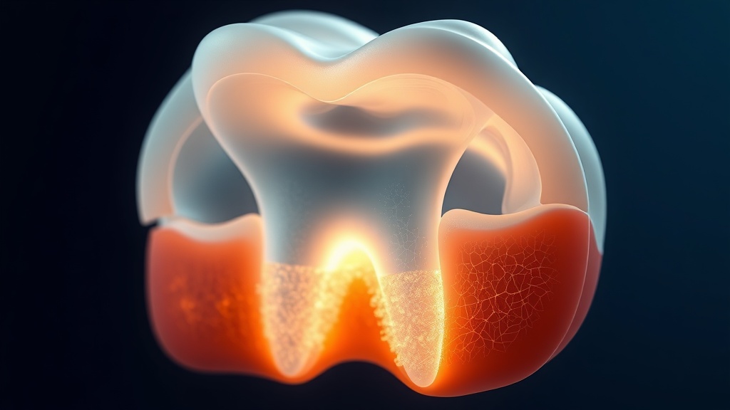
Fluorosis is built into the enamel during formation: a faint hypomineralised, porous band sits beneath the smooth outer surface.
What the research actually shows
Every claim below maps to a named, peer-reviewed source in the Sources section. According to PubMed.
| Claim | Evidence | Source |
|---|---|---|
| In recent US national survey data, most children and adolescents showed some degree of dental fluorosis, and higher fluoride exposure raised the odds of it. | Cross-sectional NHANES analysis (2013–2016). | Hung et al., 2023 |
| The large majority of dental fluorosis in the United States is very mild. | NHANES prevalence analysis (1999–2004). | Akinkugbe et al., 2014 |
| Fluorosis forms when excess childhood fluoride leaves a hypomineralised, porous enamel subsurface; severe cases add pitting and secondary brown staining. | Review of the mechanism of enamel fluorosis. | DenBesten & Li, 2011 |
| Excess fluoride acts on the enamel-forming ameloblasts, delaying protein removal so the maturing enamel stays more porous. | Review of fluoride effects on ameloblasts. | Bronckers et al., 2009 |
| Enamel microabrasion is an effective first-line cosmetic option for fluorosis marks, with high patient acceptance. | Clinical study of microabrasion for stained fluorosis. | Divyameena et al., 2021 |
How fluorosis differs from other marks
| Mark you see | What it usually is | When it forms |
|---|---|---|
| Chalky-white flecks or lacy lines | Mild dental fluorosis | During childhood tooth formation |
| White spots appearing after braces | Early demineralisation under plaque | After teeth erupt, around brackets |
| General yellowing with age | Enamel thinning as darker dentin shows through | Gradually, into adulthood |
| Brown marks on the surface | Coffee, tea, wine or tobacco on the enamel | Any time, from diet and habits |
| Brown mottling with pitting | More severe dental fluorosis | During childhood tooth formation |
Why some fluorosis is white and some is brown
Not all fluorosis looks the same, and the difference comes down to how porous the enamel became. In mild fluorosis, only the surface layers are slightly under-mineralised, so you see faint white opaque striations or small cloudy flecks — often noticeable only when the teeth are dry, and easy to miss entirely. As the degree increases, more of the enamel is affected and the surface can become pitted. Here is the part that surprises people: the brown colour seen in stronger fluorosis is usually not the fluoride itself. The porous, pitted enamel behaves like a sponge, taking up pigments from food, drinks and the environment over the years after the tooth erupts. So the white is built in during childhood, while the brown is picked up later. The timing window matters too. Because enamel fluorosis can only happen while a tooth is forming, the risk period runs from birth to roughly age 8, when the crowns of the permanent teeth are being mineralised — the lower front teeth finish earliest, around age 2 to 3, and the back teeth much later. Once those teeth are in the mouth, they cannot develop new fluorosis no matter what.
Evidence you can act on.
Occasional emails — new research, new protocols, no noise.
How the look of fluorosis can be improved
You cannot undo fluorosis, because it is built into the enamel. What you can change is how noticeable it is. None of the options below is a medical remedy for a disease — they are cosmetic ways to make the marks blend in, and the right choice depends on the severity, which a dentist grades in person.
- 1
Start with a professional assessment
one visitA dentist grades how mild or severe the fluorosis is and confirms the marks really are fluorosis rather than something else that looks similar. That grading is what decides which cosmetic option fits — and rules out anything that needs different care.
- 2
Enamel microabrasion — a gentle surface polish
usually one visitFor mild surface white and brown marks, a dentist can remove an extremely thin outer layer of enamel using a fine abrasive paste with a mild acid. In clinical studies it is an effective, well-accepted first-line option for fluorosis marks, and it is conservative because it touches only the very surface.
- 3
Resin infiltration — filling the pores optically
usually one visitA low-viscosity resin is drawn into the porous enamel so it bends light like healthy enamel, and the white simply blends in. In a systematic review it masked marks significantly better than natural remineralisation or fluoride varnish, and one trial found it more effective than microabrasion for fluorotic white spots. There is no drilling.
- 4
Whitening — with honest expectations
days to weeksWhitening the whole tooth can even out the contrast for some people, but it does not remove fluorosis, and it can briefly make white spots look more obvious before the surrounding enamel lightens. In one study, pairing microabrasion with in-office whitening improved appearance more than microabrasion alone. Think of whitening as a finishing step, not the fix.
- 5
For severe cases, ask about covering options
planned with a dentistWhen pitting and brown mottling are extensive, a dentist may suggest bonding or veneers that cover the enamel surface entirely. This is a bigger, planned cosmetic decision made in person, not something to attempt at home.
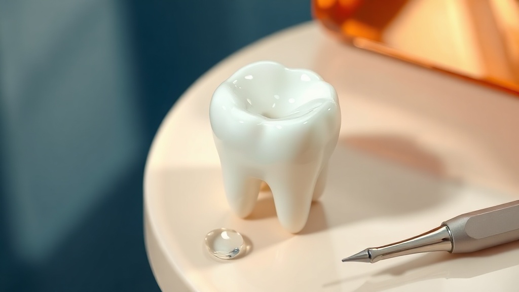
Minimally-invasive options like microabrasion and resin infiltration aim to make fluorosis marks blend in, not to strip the tooth.
See a dentist if the marks involve brown mottling or visible pitting, if you are not certain the discolouration is fluorosis, or if you want to improve how mild fluorosis looks — microabrasion and resin infiltration are dentist-applied procedures. Any questions about a child's fluoride intake are best answered by a dentist or doctor, who can match the right amount to the child's age.
Frequently asked questions
Sources
- 1.
- 2.
- 3.
- 4.
- 5.
- 6.
- 7.
- 8.
- 9.
- 10.
- 11.
- 12.

Fix your breath at the source.
The complete science-backed protocol — engineered to eliminate volatile sulfur compounds at the biological source.
Start the Breath Protocol →Related reading
Educational purposes only. The content on this page is not medical advice and is not a substitute for consultation with a qualified dental or medical professional.
More from the library
 Causes8 min
White Spots on Teeth After Braces: Causes and How to Fix Them
What causes the white marks left after braces come off, and the evidence-based cosmetic options that make them fade or blend in.
Read →→ Causes8 min
Brown Stains on Teeth: What Causes Them and How to Fade Them Safely
Most brown tooth stains are surface deposits from everyday food, drink and tobacco. Here is why they form and the enamel-safe ways to lighten them.
Read →→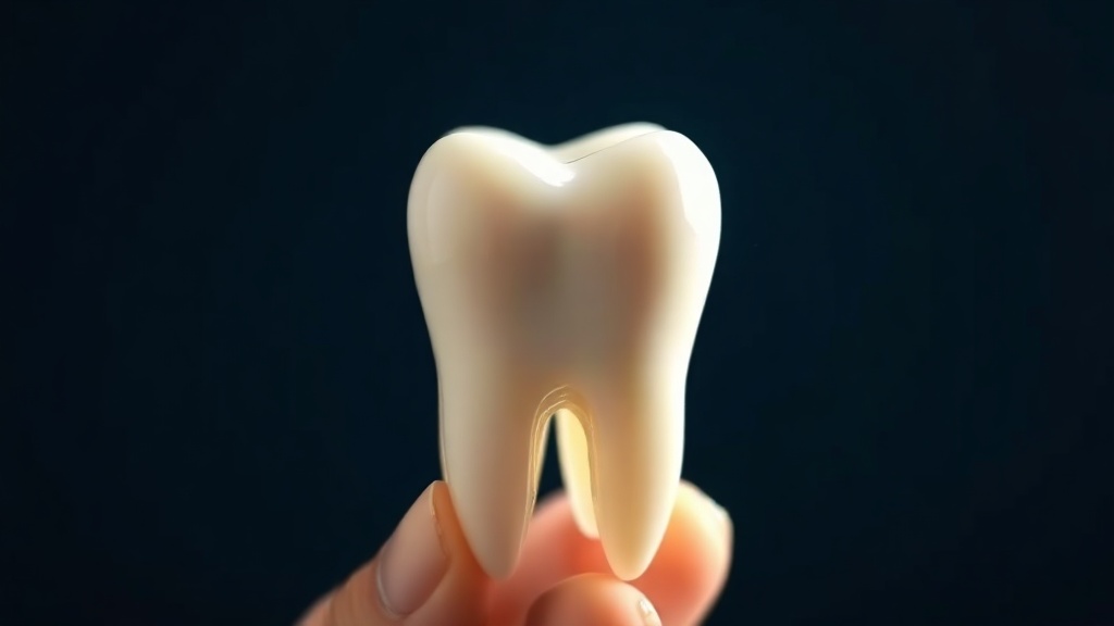 Answers8 min
Does Teeth Whitening Damage Enamel? The Evidence-Based Answer
The honest, evidence-based answer to whether whitening harms enamel — what peroxide really does to the surface, why sensitivity is not damage, and the DIY methods that actually cause wear.
Read →→ Guides8 min
How to Get Rid of Yellow Teeth: An Enamel-Safe Guide
Two kinds of yellow, two different fixes, and the enamel-safe way to brighten each one.
Read →→ Answers8 min
Does Baking Soda Whiten Teeth? What the Evidence Says
An honest look at what baking soda can and cannot do for a whiter smile.
Read →→ Best Of9 min
Best Fluoride-Free Whitening Toothpaste: An Honest, Evidence-Based Guide (2026)
Which fluoride-free whitening ingredients hold up to the research — and which are marketing. An honest buyer's guide.
Read →→